In oxygen (O2) distribution in tissue, two stages can be discerned:
release from the erythrocyte's
hemoglobin (Hb)
up to the capillary
border, and from there, diffusion into the tissue. The latter is handled under
Tissue Oxygenation.
The release of O2 from Hb is no problem, but after that, the O2 has to be transported further through blood and capillary wall, and the consequence is, that the surrounding tissue 'sees' an O2 partial pressure PO2 that is lower than the erythrocyte's PO2. The diffence is called Extraction Pressure (EP) (1).
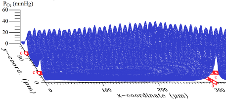
The figure shows a computer model calculation for a hard working muscle, alongside two capillaries (c) supplied by an arteriole (a). The peaks represent PO2 in the erythrocytes; about the top ¼ of the peak height is within the capillary, the bottom ¾ is in the surrounding tissue.
Calculating the EP is not easy and requires an elaborate computer program (2). However, from these computer calculations an approximate formula for human blood was derived together with source lines in the computer language C. A summary is in this document – or consult chapter 9 of the dissertation, via https://repository.ubn.ru.nl/handle/2066/146333